New Developments
A new technology has recently been developed and tested for simultaneous recordings of electrical activity (electromyograms, EMGs) in multiple Manduca muscles.
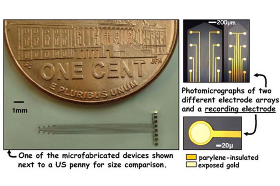
Working with Prof. Robert White (Mech. Eng) in the Tufts Micro and Nano Fabrication facility, PhD graduate student Cinzia Metallo (Neuroscience, Sackler School) has succeeded in designing, fabricating and testing flexible multi-electrode arrays that can be implanted into freely moving caterpillars to monitor the neural patterns controlling behavior. The electrode arrays consist of a chrome/gold conductive layer sandwiched between two insulating layers of Parylene C. One of the micro-fabricated devices is shown below.
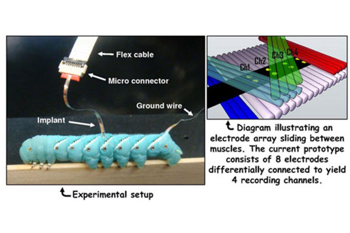
To ensure ease of insertion, optimal placement, and recording stability, the geometry of the electrode array has been designed to match the muscle anatomy of Manduca. When the electrode arrays are implanted between muscles layers and connected to an extracellular amplifier, the individual electrical pulses associated with nerve stimuli can be resolved at multiple locations. The typical experimental setup is shown in the figure below.
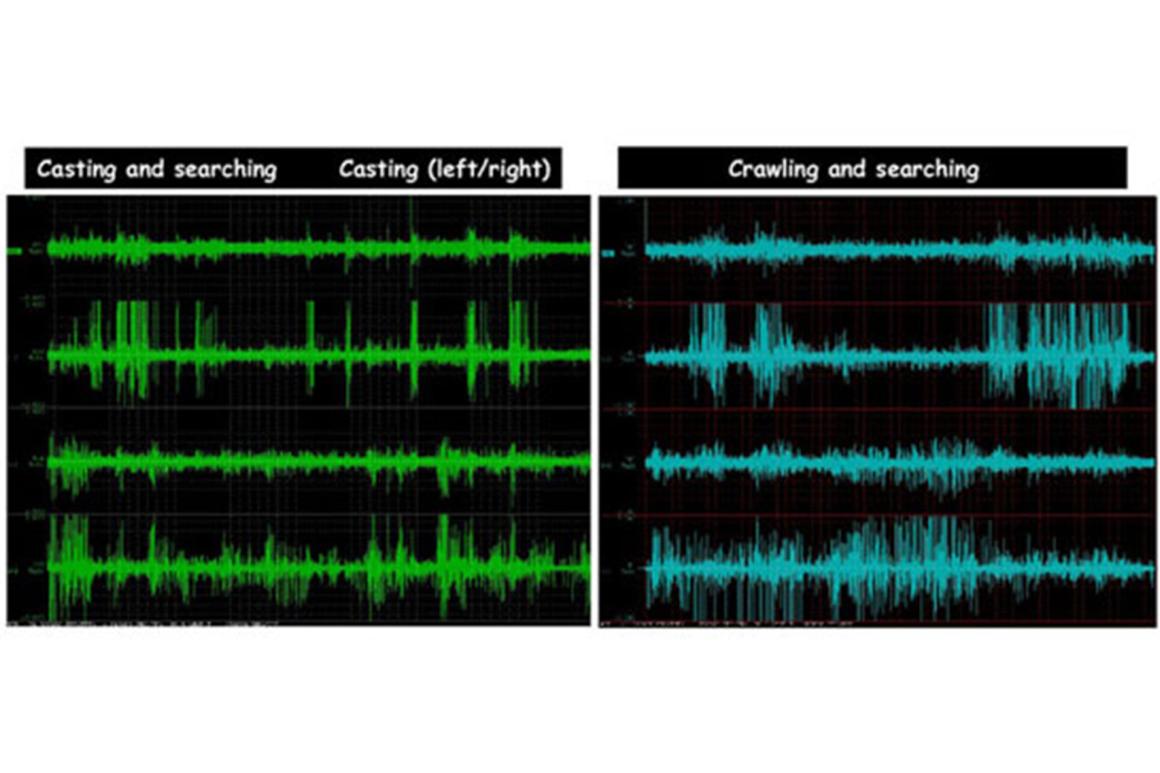
Because larval Manduca muscles are generally innervated by a single motor neuron, the EMGs can be resolved into spikes representing the patterns of neural commands by identified neurons. The arrays are now being used to identify activity in different neurons during crawling, climbing, turning and searching behaviors. This is a significant technological advancement that will greatly accelerate our understanding of neuromechanical control of soft animal locomotion. Examples of four-channel EMG recordings taken during different behaviors are shown below.